
口ゴボの治療計画と結果|歯・骨格・口唇のタイプ別に、論文から読み解く
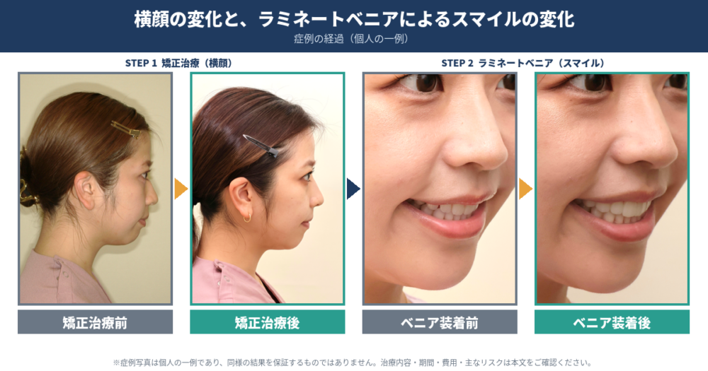
トップの画像は当院の上村先生の抜歯矯正前後の比較です。矯正治療だけでなぜか鼻が高くなったような変化。べニア治療とオトガイ下制筋へのボツリヌス注射も経て魅力的なスマイルへ。この変化こそ中嶋プロデュース(笑)
この記事の出典・データソース
本記事は、口ゴボ(上下顎前突・上下顎歯槽性前突)の矯正治療に関する約50編の学術論文(系統的レビュー、専門家合意、後ろ向き・前向き研究、症例報告)を、AI文献検索エンジン「Consensus」で整理した内容をもとに、矯正歯科医が監修・加筆したものです。引用した論文は記事末尾の参考文献(DOIリンク付き)に掲載しています。論文の結果は研究対象の集団・条件での平均的な傾向であり、個々の患者さんに同じ結果を保証するものではありません。
この記事のポイント
- 「口ゴボ」は見た目を表す言葉で、前歯の傾き・顎の位置関係・口唇などの軟組織が組み合わさって生じます。原因によって治療の考え方が変わります。
- 小臼歯抜歯を伴う矯正で口唇の突出が減る方向は、研究間でおおむね一致しています。ただし前歯が動いた量と、口唇が動く量は同じではなく、個人差が大きいことが分かっています。
- 後退量には歯槽骨の厚みや歯根吸収といった解剖学的な安全範囲があり、「できるだけ下げる」ことが目標ではありません。
- 重い骨格的な不調和には外科的矯正治療という選択肢がありますが、合併症や後戻りも含めて説明を受けて選ぶことが大切です。
口ゴボとは?見た目の言葉であり、原因は一つではありません
「口ゴボ」は、横顔で口元が前に出て見える状態を指す日常的な言葉です。医学的には、上下の前歯が前に傾いている状態(上下顎歯槽性前突)や、上下の顎の位置関係が関わる状態(上下顎前突)などを含みます。
研究では、突出感を作る要因として次のものが挙げられています (Pan et al., 2025)。
- 上下の前歯の前方への傾き
- 上顎と下顎の前後的な位置関係(下顎が小さい・後ろに下がっている場合など)
- 顔の縦方向の長さ
- 口唇の厚みや緊張といった軟組織の状態
つまり、「口ゴボ」と一言でいっても、前歯の傾きが中心の方もいれば、骨格の影響が大きい方もいます。ここを見分けることが、治療計画の出発点です。
適切に選んだ歯の移動で前歯を後退させると口唇の突出は改善する、というのが研究の大きな流れですが、変化量には個人差があり、重い骨格的不調和では外科的矯正治療も検討されます (Bills et al., 2005; Leonardi et al., 2010; Pan et al., 2025)。
また、治療の成功は横顔のラインだけでは評価できません。研究で目標とされるのは、適正な前歯の傾き、正常な前歯のかみ合わせ、安定した咬合、口唇が自然に閉じられること、そして患者さんが望む顔立ちとの調和です (Huang et al., 2022; Jawale et al., 2021)。
診断:治療方針を決める前に何を調べるのか
治療方針は、精密検査の結果をもとに組み立てます。論文では、次のような検査を組み合わせて評価するとされています (Chu et al., 2009; Pan et al., 2025)。
- 顔貌・口腔内の診察:口を閉じやすいか、笑ったときの歯ぐきの見え方、歯の見え方など
- 側面頭部X線規格写真(セファログラム):顎の位置や前歯の傾きを数値で評価
- パノラマX線・歯列模型:歯の本数や位置、叢生(歯並びのでこぼこ)、開咬・過蓋咬合の有無
- 口唇の評価:安静時に閉じやすいか、閉じたときにオトガイ(あご先)の筋肉が緊張しないか
- 歯周組織・顎関節・気道・筋機能・成長段階の確認
必要に応じて歯科用コーンビームCT(CBCT)で歯根と骨の位置関係を調べることもありますが、撮影は被曝を抑える原則に従い、必要性を個別に判断して行います (Pan et al., 2025)。
当院では、骨格や歯の位置だけでなく、上顎前歯の三次元的な位置を起点に、顔貌との調和を設計する考え方(Face-Driven Dentistry:FDD)を診断の軸にしています。横顔の「口元だけ」を見るのではなく、顔全体の中で前歯がどこにあるべきかを検討します。
治療の選択肢:歯の移動、垂直的な制御、外科的矯正治療
研究で報告されている主な方針を、状態別に整理します。どの方法が適するかは精密検査をもとに矯正歯科医が個別に判断するもので、この表は一般的な研究の整理です。
| 主な状態 | 研究で用いられる方針 | 計画上の注意 |
|---|---|---|
| 前歯の前傾・突出が中心 | 歯の移動による前歯の後退 | 必要な空隙の量と、口唇の変化を評価する (Chu et al., 2009) |
| 軽度・境界的な突出 | 歯列全体を後方へ動かす方法など | 他の方法との比較根拠は限定的 (Sanman et al., 2025) |
| 顔が縦に長い・歯ぐきの露出を伴う | 歯を沈める(圧下)など垂直的な制御 | 顔貌と歯の露出量に応じて設計する (Pan et al., 2025) |
| 重い骨格的不調和 | 外科手術と、術前・術後の矯正の併用 | 歯の移動だけで補える限界を評価する (Pan et al., 2025) |
【根拠は弱い〜傾向】 歯列全体を後方へ動かす方法は、小規模な比較研究で、抜歯を伴う方法より変化量がやや小さい傾向が示されています。境界症例26人を調べた研究も、治療選択と歯列弓の形の関連を記述したにとどまり、選択基準の確立には至っていません (Sanman et al., 2025; Gautam et al., 2026)。
成長期のお子さんでは、下顎の成長を誘導する方法が検討されます。一方で、顔が縦に長く、口唇を閉じるときの筋肉の緊張が強い場合は、機能的な装置が向かないことがあります。成人では、成長誘導を前提とせず、歯槽性の補償と外科的な修正を区別して考えます (Pan et al., 2025)。
なお、どの方法を選ぶか(抜歯の要否を含む)は、突出感だけでは決まりません。突出の程度、叢生、垂直的な問題、歯周組織の状態などを総合して判断するため、この記事では一律の基準はお示ししません。
期待できる変化:口唇はどれくらい動くのか
変化の方向は一致しています
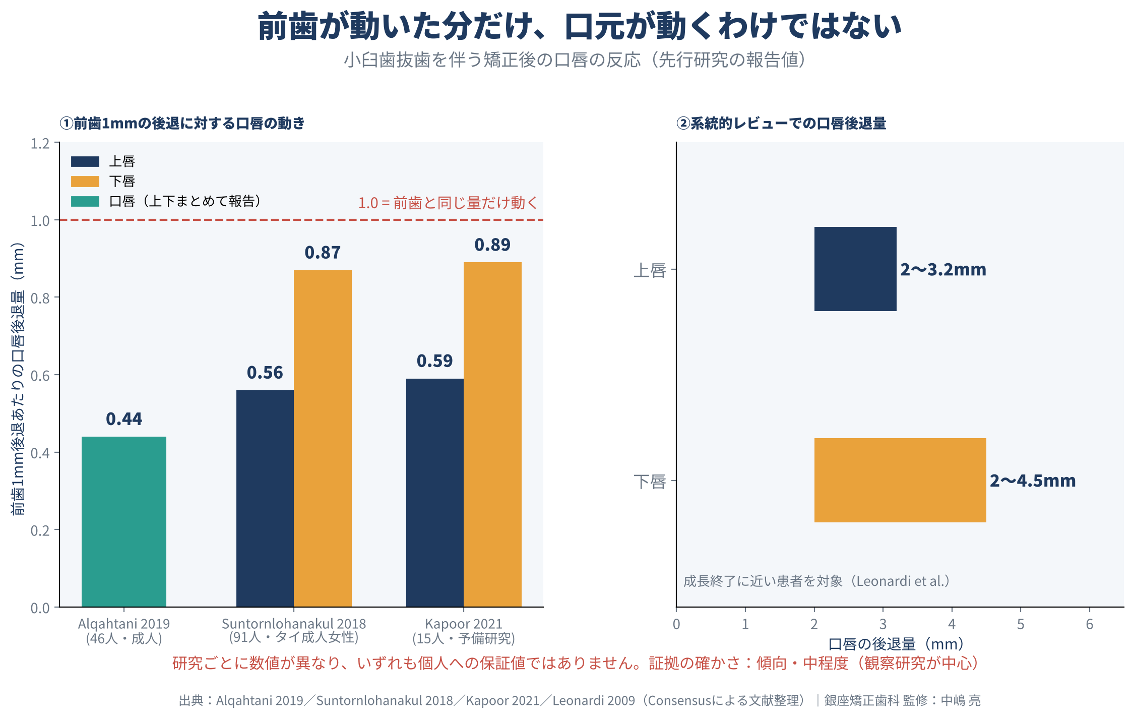
【傾向・中程度の根拠】 抜歯を伴う矯正治療の後は、前歯が後退して立ち上がり、口唇の突出が減り、鼻と上唇の角度(鼻唇角)が大きくなる、という方向の変化が複数の研究で一致して報告されています。系統的レビューでは、成長終了に近い患者で上唇が約2〜3.2mm、下唇が約2〜4.5mm後退したとされています (Leonardi et al., 2010; Alqahtani et al., 2019; Mohapatra et al., 2024; Agarwal et al., 2023; A et al., 2024)。ただしこの根拠の多くは観察研究(治療前後の比較)です。第二小臼歯を抜歯した成人女性の後ろ向き研究でも軟組織の改善は報告されていますが、下顎前歯に有意な変化がない項目もありました (Alqahtani et al., 2020)。
前歯1mmに対して、口唇は1mm動くわけではありません
ここで注意したいのは、「改善する」という結論と、「何mm変わるかを個人単位で予測できる」という結論は別だということです。系統的レビューでも、反応の大きな個人差が指摘されています (Elias et al., 2023; Leonardi et al., 2010)。
- 成人46人の研究:前歯1mmの後退に対し、口唇は約0.44mm (Alqahtani et al., 2019)
- タイの成人女性91人の研究:上顎前歯と上唇が1:0.56、下顎前歯と下唇が1:0.87 (Suntornlohanakul et al., 2018)
- 15人の予備的な研究:上唇0.59mm、下唇0.89mm (Kapoor et al., 2021)
研究ごとに推定値が異なり、いずれも個人への保証値ではありません。
| 評価項目 | 報告された数値・結果 | 解釈 |
|---|---|---|
| 口唇の後退量 | 上唇2〜3.2mm、下唇2〜4.5mm | 成長終了に近い患者の系統的レビュー (Leonardi et al., 2010) |
| 前歯の変化 | 平均約4.1mm後退、上下約9.6°直立化 | 成人46人の後ろ向き研究 (Alqahtani et al., 2019) |
| 三次元的な軟組織の変化 | 正中以外でも2〜4mm後退 | 成人アジア人24人の研究 (Solem et al., 2013) |
| 下顔面の高さ | 有意な変化なし | 後退だけで顔が短くなるとは限らない (Harahap & Oeripto, 2019) |
口唇の厚みや形でも反応は変わります
口唇の厚みと反応の関係も一定ではありません。成人75人の研究では厚い口唇ほど後退量が小さい一方、15人の研究では厚い上唇でより大きな後退と関連したと報告されています。単一の指標だけで結果を予測するのには限界があります (Albertini et al., 2024; Veni et al., 2023)。日本人の成人女性33人の研究でも、前歯の位置、咬合平面、治療前の口唇の隙間など、複数の要素が口唇の変化に関わっていました (Hayashida et al., 2011)。
【根拠は弱い(症例報告)】 口が閉じやすくなり、オトガイの筋肉の緊張が減ったという結果は、症例報告で繰り返し示されています。ただし全員に同じ改善が起こるわけではなく、重い骨格性の上顎前突(Ⅱ級)の症例では、横顔が改善しても口唇が閉じにくい状態が残った報告もあります (Huang et al., 2022; Liaw et al., 2025; Chen et al., 2025)。
治療期間の目安:症例によって大きく異なります
治療期間も、研究によって幅があります。上下顎前突38人を対象にした研究では26±4か月でした (Mao et al., 2019)。症例報告では16か月、18か月、27か月という例があり、マウスピース型矯正装置を用いた複合的な開咬症例では33か月でした。欠損歯への対応などが加わる症例では、35〜48か月を要した報告があります (Lishoy et al., 2020; Halwa et al., 2019; Huang et al., 2022; Xiang et al., 2023; Yu et al., 2023; Deng et al., 2024)。
外科的矯正治療を含む185人の調査では、総治療期間の中央値は31.1か月でした (Paunonen et al., 2017)。手術そのものの回復期間と、治療全体の期間は区別して考える必要があります。
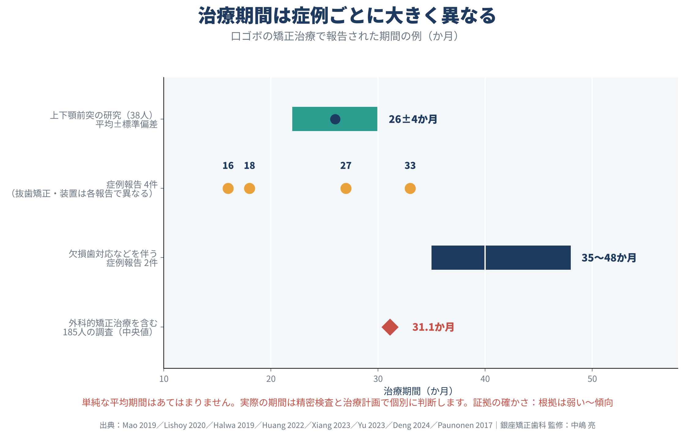
こうした報告から、単純な平均期間を一人ひとりにあてはめることは適切ではありません。実際の期間は、精密検査と治療計画をもとに個別にお伝えします。
後退量の限界と安全性:「できるだけ下げる」は目標ではありません
矯正用アンカースクリュー(TADs)の役割
矯正用アンカースクリュー(temporary anchorage devices:TADs)は、奥歯が意図せず前に動くのを抑え、前歯の後退や垂直的な制御に使われます。口元とかみ合わせの改善を示す報告がありますが、装置が使えることと、安全に大きく動かせることは同じではありません (Malhotra et al., 2024; Vishnani et al., 2024; Liu et al., 2021)。
歯槽骨の厚み
【傾向・中程度の根拠】 重要な制約は、歯根を囲む歯槽骨です。前歯を後退させると、口蓋側(上あご)や舌側(下あご)の骨が薄くなり、骨裂開(骨が部分的に失われる状態)が生じることがあります。二次元のX線や通常の診察では把握しにくい場合もあります (Sarikaya et al., 2002; Nayak et al., 2013)。三次元研究では、もともと薄く、骨の入れ替わりが小さい口蓋側の歯槽骨の上縁部への注意が強調されています (Ito et al., 2023)。
成人は若年者よりも骨の支えの減少が大きい傾向があり、歯周炎を伴う方では口蓋側の骨の減少と骨裂開がより多く認められました (Zheng et al., 2022; Li et al., 2025)。骨の変化は歯の移動量に一対一で対応するわけではなく、後退量だけで骨の反応を予測するのも困難です (Hong et al., 2019)。
歯根吸収
【傾向・中程度の根拠】 歯根吸収(歯の根の先が短くなる変化)も無視できません。成人36人の研究では、大きな後退・圧下と歯根吸収が関連していました。40人を解析したランダム化試験では、骨に固定したスクリューによる圧下方向の力が、前歯の吸収リスクを高める結果でした (Chen & Liu, 2020; Arvind et al., 2023)。37人を対象とした別の研究では、上顎の切歯の歯根が平均0.81±0.46mm短くなり、成人32人の前向き研究でも下顎切歯で歯根長の減少が報告されています (Wang et al., 2018; Prajapati et al., 2025)。
口唇の平坦化と気道
顔貌の面でも、後退しすぎると口唇が平坦になり、横顔が不自然になることがあります。突出を減らすことと、口唇のふくらみを残すことのバランスが重要です (Pan et al., 2025; Chu et al., 2009)。
また、前歯の後退後に気道が狭くなる可能性は専門家の合意文書でも検討されていますが、提示された根拠だけから「必ず呼吸の問題が起こる」とは判断できません。すでに気道が狭い方では、後退量を計画する際の配慮事項とされています (Pan et al., 2025)。
外科的矯正治療と長期安定性
外科的矯正治療とは
重い骨格的不調和がある場合は、外科手術と矯正の併用が選択肢になります。前歯部の歯槽骨切り術、Le Fort I型骨切り術、下顎骨切り術などがあり、必要に応じて組み合わせます。前歯部だけの突出か、顎全体の位置や縦方向の過剰を伴うかによって設計が異なります (Chu et al., 2009)。
51人の後ろ向き研究では、上下顎の前歯部の歯槽骨切り術を併用した患者で、目標とした前歯の角度に到達しやすく、治療期間も短縮されたと報告されました。ただし比較対象も顎矯正手術を受ける患者であり、矯正単独の症例との単純な比較ではありません (Yong et al., 2022)。
安全性:一律に「少ない」とは言えません
| 長期評価・安全性 | 研究結果 | 読み取り方 |
|---|---|---|
| Le Fort I型骨切り術(分割)の術後 | 2年で前歯の傾きが平均2.1〜2.6°後戻り | 小さな変化と治療の失敗は区別して考える (Tai et al., 2022a) |
| 下顎の前歯部骨切り術の術後 | 2年で平均2.9°後戻り | 著者らは臨床的な影響は小さいと評価 (Tai et al., 2022b) |
| 術後の合併症 | 創の離開、感染、歯肉退縮、歯周の骨減少など | 術式・施設・評価方法で頻度が異なる (Tai et al., 2022a) |
| 縦に長い骨格性Ⅱ級 | 2mmを超える後戻りが44%、低〜中角群では20% | 成人52人の単施設研究 (Poolsin et al., 2025) |
| 抜歯矯正後の骨 | 保定中に部分的に回復 | 治療前の状態まで完全に戻るとは限らない (Wang et al., 2023) |
手術の安全性を一律に「合併症は少ない」と表現するのは適切ではありません。20例の前向き研究では歯の神経が失われた(失活歯)例があり、系統的レビューでも感染、隣の歯の損傷、瘻孔などが報告されています。一方、262人の別の手術コホートでは歯肉退縮や歯髄の損傷の頻度は低く、施設によって結果は大きく異なります (Gupta et al., 2019; Haas et al., 2017; Posnick et al., 2016)。
下顎の骨切りを含む手術では、下唇の感覚の低下が残る方がいることも重要な説明事項です。生命に関わる合併症はまれでも、周術期・術後の問題全体は珍しくありません (Thiem et al., 2020; Kotaniemi et al., 2023)。
手術を先に行う「サージェリーファースト」は、早期の顔貌改善と期間の短縮が利点とされますが、適応と長期安定性の根拠は十分ではありません (Pan et al., 2025)。
保定と後戻り
【傾向・中程度の根拠】 矯正後の保定(歯並びを安定させる期間)の研究では、若年34人で18〜24か月間に明らかな前歯の後戻りがなかった一方、成人の骨欠損の追跡では、骨の回復とともに前歯のわずかな向きの変化も認められました (Wang et al., 2021; Guo et al., 2023)。5年間安定した症例報告もありますが、一般的な境界症例の長期比較では、抜歯・非抜歯のどちらにも前歯の不整な再発があり、抜歯をしたから後戻りしない、とは言えません (Deng et al., 2024; Aras et al., 2025)。
外科治療の長期メタ解析では、多くの平均変化が臨床的に許容できる範囲内でしたが、評価方法や対象のばらつきが大きく、安定性を断定することには限界があります (Gaitan-Romero et al., 2020; Eckmüller et al., 2022)。
裏側矯正と口ゴボ:当院での考え方
口ゴボの治療でも、装置を歯の裏側に付ける裏側(舌側)矯正を選択される方がいらっしゃいます。論文では、デジタル設計の舌側装置とマイクロスクリューを併用して、骨格性の上下顎前突とガミースマイルを治療した症例が報告されています (Chen et al., 2023)。ただしこれは【根拠は弱い】症例報告であり、治療方法の優劣を示すものではありません。
当院では、装置の種類を選ぶ前に、先ほど述べたような精密検査で「何が突出感をつくっているのか」を見極め、骨や歯根の安全範囲を確認したうえで、顔貌との調和を設計します。ガミースマイルを伴う場合の考え方はガミースマイルの原因別治療法で、成人矯正で歯の移動だけでは補えない限界については成人矯正の限界と治療ゴールで詳しく解説しています。口元の美しさの指標として知られるEラインについてはEラインと矯正治療をご覧ください。
装置については、裏側矯正(舌側矯正)とマウスピース矯正のページで特徴を紹介しています。費用は料金のご案内をご確認ください。矯正歯科医の選び方に迷われる方は、矯正歯科の医院選びガイドや認定医についての解説も参考になります。
治療のリスク・副作用について
矯正治療には、一般に次のようなリスク・副作用があります。治療前に十分にご説明します。
- 歯の移動に伴う痛みや違和感、装置による口内炎
- 歯根吸収や、歯槽骨の菲薄化・骨裂開、歯肉退縮
- むし歯・歯周病のリスク(清掃が難しくなるため)
- 後戻り(保定装置を指示どおり使わない場合など)
- 外科的矯正治療を併用する場合は、感覚の麻痺、感染などの手術に伴うリスク
よくあるご質問
Q. 口ゴボは矯正治療で必ず改善しますか?
A. 研究では、口唇の突出が減る方向で結果は一致していますが、改善量には個人差が大きく、改善を保証することはできません。骨格の影響が大きい場合は、外科的矯正治療が検討されることもあります。
Q. 前歯を下げた分だけ、口元も下がりますか?
A. 同じ量にはなりません。研究では、前歯1mmの後退に対して口唇は約0.4〜0.9mmと幅があり、下唇のほうが上唇より動きやすいと報告されています。
Q. 治療期間はどれくらいですか?
A. 報告では16か月から48か月まで幅があります。精密検査のうえで、個別にお伝えします。
Q. できるだけ口元を下げたほうがいいのでしょうか?
A. そうとは限りません。後退しすぎると、骨が薄くなる、歯根が短くなる、口唇が平坦になるといった問題が起こることがあります。安全な範囲の中で、顔全体の調和を目指します。
まとめ
- 口ゴボの治療は、前歯の傾き・骨格・口唇の状態を精密検査で見極めたうえで、個別に設計されます。
- 口唇の突出が減る方向の変化は研究間で一致していますが、変化量は個人差が大きく、前歯が動いた量と口唇の動きは一致しません。
- 後退量には、歯槽骨の厚みや歯根吸収などの安全範囲があります。
- 外科的矯正治療は選択肢の一つですが、合併症や後戻りについても理解して選ぶことが大切です。
- 近年の流れは、「口元をできるだけ下げる」治療から、歯・骨格・口唇と安全限界を統合する個別計画へ移っています。個人の見た目の改善量と長期安定性をどこまで正確に予測できるかは、今後の課題です。
ご自身の口元が気になる方は、まず精密検査で現状を知ることから始めてみませんか。無料相談のご予約はこちらから承っています。
監修者
中嶋 亮(なかじま りょう)
日本デジタル矯正歯科学会理事/銀座矯正歯科院長/医療法人社団真美会理事長
日本矯正歯科学会認定医、ESLO(ヨーロッパ舌側矯正歯科学会)Titular member
詳しい経歴は院長・スタッフ紹介をご覧ください。
本記事は一般的な情報提供を目的としており、個別の診断・治療を代替するものではありません。治療の可否や方法は、精密検査のうえ担当の矯正歯科医にご相談ください。
記事公開日:2026年11月5日/最終更新日:2026年11月5日
参考文献
DOIは文献検索(Consensus)の出力に基づきます。
A, A. Y., Chundawat, M. S., Sambhwani, J., Anjum, S., & Felazin, M. (2024). Changes in the curvature of upper and lower lips following the first premolar extraction in Class I bimaxillary protrusion: A cephalometric study. IP Indian Journal of Orthodontics and Dentofacial Research.
Agarwal, P., Hoshing, S., & Shinde, S. (2023). Evaluating the effects of first premolar extraction on Point A, Point B and nasolabial angle in patients with bimaxillary protrusion. Journal of Medical Science and Clinical Research. https://doi.org/10.18535/jmscr/v11i3.04
Albertini, P., Barbara, L., Albertini, E., Willeit, P., & Lombardo, L. (2024). Soft-tissue profile changes in adult patients treated with premolar extractions. American Journal of Orthodontics and Dentofacial Orthopedics. https://doi.org/10.1016/j.ajodo.2024.04.011
Alqahtani, N., Alqasir, A., Al-Jewair, T., Almoammar, K., & Albarakati, S. F. (2020). Dental and soft tissue changes following extraction of second premolars in females with bimaxillary protrusion: A retrospective study. Nigerian Journal of Clinical Practice, 23, 1110–1119. https://doi.org/10.4103/njcp.njcp_636_19
Alqahtani, N., Alshammari, R., Almoammar, K., Almosa, N., Almahdy, A., & Albarakati, S. F. (2019). Post-orthodontic cephalometric variations in bimaxillary protrusion cases managed by premolar extraction: A retrospective study. Nigerian Journal of Clinical Practice, 22, 1530–1538. https://doi.org/10.4103/njcp.njcp_125_19
Aras, I., Griffith, R. R., Trouten, J., Alexander, M., & Akyalcin, S. (2025). Long-term relapse of anterior teeth in orthodontic patients treated with and without premolar extractions using a 3-dimensional surface mesh analysis. American Journal of Orthodontics and Dentofacial Orthopedics. https://doi.org/10.1016/j.ajodo.2025.02.020
Arvind, T. P. P., Ramasamy, N., Subramanian, A., Selvaraj, A., & Siva, S. (2023). Three-dimensional volumetric evaluation of root resorption in maxillary anteriors following en-masse retraction with varying force vectors: A randomized control trial. Orthodontics & Craniofacial Research. https://doi.org/10.1111/ocr.12704
Bills, D., Handelman, C., & BeGole, E. (2005). Bimaxillary dentoalveolar protrusion: Traits and orthodontic correction. The Angle Orthodontist, 75(3), 333–339. https://doi.org/10.1043/0003-3219(2005)75[333:bdptao]2.0.co;2
Chen, Y.-H., Gao, Y., He, J.-J., Fu, S.-M., & Wang, L. (2023). Digital lingual appliance combined with micro-screws for the treatment of a skeletal bimaxillary protrusion and ‘gummy’ smile. Australasian Orthodontic Journal, 39, 96–108. https://doi.org/10.2478/aoj-2023-0011
Chen, Y., & Liu, D.-X. (2020). Morphologic evaluation of root resorption after miniscrew assisted en mass retraction in adult bialveolar protrusion patients. Head & Face Medicine, 16. https://doi.org/10.1186/s13005-020-00229-z
Chen, Z.-X., He, J., Wang, L.-F., Gan, X.-P., Zhong, P., & Mo, S.-X. (2025). Alveolar bone dehiscence in anterior teeth after orthodontic camouflage treatment for severe skeletal Class II malocclusion with a history of condylar resorption at the 3-year follow-up: A case report. BMC Oral Health, 25. https://doi.org/10.1186/s12903-025-05658-3
Chu, Y.-M., Bergeron, L., & Chen, Y.-R. (2009). Bimaxillary protrusion: An overview of the surgical-orthodontic treatment. Seminars in Plastic Surgery, 23, 32–39. https://doi.org/10.1055/s-0028-1110099
Deng, J., Zeng, S., Chen, Y.-J., Shao, Q., & Zhou, S. (2024). A patient with bimaxillary protrusion was treated by the extraction of four premolars and four compromised first molars: A case report. Australasian Orthodontic Journal, 40, 37–49. https://doi.org/10.2478/aoj-2024-0001
Eckmüller, S., Paddenberg, E., Hiller, K., Proff, P., Knüttel, H., & Kirschneck, C. (2022). Relapse in class II orthognathic surgery: A systematic review. BMC Oral Health, 22. https://doi.org/10.1186/s12903-022-02636-x
Elias, K. G., Sivamurthy, G., & Bearn, D. (2023). Extraction vs nonextraction orthodontic treatment: A systematic review and meta-analysis. The Angle Orthodontist. https://doi.org/10.2319/021123-98.1
Gaitan-Romero, L., Shujaat, S., H., Orhan, K., Shaheen, E., Mulier, D., Willems, G., Politis, C., & Jacobs, R. (2020). Evaluation of long-term hard tissue relapse following surgical-orthodontic treatment in skeletal class II patients: A systematic review and meta-analysis. International Journal of Oral and Maxillofacial Surgery. https://doi.org/10.1016/j.ijom.2020.09.001
Gautam, R., Ahuja, D., Shetty, S., Ashith, M., Batra, P., Chandhini, G., Khera, A., Mithra, P., & Shenoy, N. (2026). Assessment of pre-treatment diagnostic characteristics in patients with borderline bimaxillary protrusion: A pilot comparison of bi-jaw distalization versus premolar extraction. International Orthodontics, 24(2S), 101134. https://doi.org/10.1016/j.ortho.2025.101134
Guo, R., Li, L., Lin, Y., Huang, Y.-P., Liu, J., Pan, M.-Q., Xu, L., & Li, W.-R. (2023). Long-term bone remodeling of maxillary anterior teeth with post-treatment alveolar bone defect in adult patients with maxillary protrusion: A prospective follow-up study. Progress in Orthodontics, 24. https://doi.org/10.1186/s40510-023-00489-w
Gupta, A., Sharma, S. D., Kataria, V., Bansal, P., & Sharma, R. (2019). Experience with anterior maxillary osteotomy techniques: A prospective study of 20 cases. Journal of Maxillofacial and Oral Surgery, 19, 119–124. https://doi.org/10.1007/s12663-019-01234-9
Haas, O., Guijarro-Martínez, R., De Sousa Gil, A. P., Da Silva Meirelles, L., De Oliveira, R. D., & Hernández-Alfaro, F. (2017). Stability and surgical complications in segmental Le Fort I osteotomy: A systematic review. International Journal of Oral and Maxillofacial Surgery, 46(9), 1071–1087. https://doi.org/10.1016/j.ijom.2017.05.011
Halwa, H. K., Yadav, S., Dutta, K., Gupta, S., Shrestha, R., & Shah, A. (2019). Bimaxillary protrusion: A case report. Journal of Universal College of Medical Sciences. https://doi.org/10.3126/jucms.v7i1.24697
Harahap, N., & Oeripto, A. (2019). Lower facial height and soft tissue changes in bimaxillary protrusion cases. Scientific Dental Journal, 3, 1–7. https://doi.org/10.26912/sdj.v3i1.3633
Hayashida, H., Ioi, H., Nakata, S., Takahashi, I., & Counts, A. (2011). Effects of retraction of anterior teeth and initial soft tissue variables on lip changes in Japanese adults. European Journal of Orthodontics, 33(4), 419–426. https://doi.org/10.1093/ejo/cjq095
Hong, S. Y., Shin, J., Hong, C., Chan, V., Baik, U., Kim, Y. H., & Chae, H. (2019). Alveolar bone remodeling during maxillary incisor intrusion and retraction. Progress in Orthodontics, 20. https://doi.org/10.1186/s40510-019-0300-2
Huang, Y.-W., Kuo, C.-L., Liu, I.-H., Tsai, Y., Wang, C.-L., & Yang, C.-H. (2022). Orthodontic treatment of severe bimaxillary dentoalveolar protrusion with skeletal Class II malocclusion without using miniscrews. Taiwanese Journal of Orthodontics. https://doi.org/10.38209/2708-2636.1121
Ito, A., Mayama, A., Oyanagi, T., Ogura, H., Seiryu, M., Fukunaga, T., Kitaura, H., & Mizoguchi, I. (2023). Three-dimensional morphologic analysis of the maxillary alveolar bone after anterior tooth retraction with temporary anchorage devices. The Angle Orthodontist, 93(6), 667–674. https://doi.org/10.2319/120122-827.1
Jawale, B., Rodrigues, L., Belludi, A., Hattarki, R. S., & Ashtekar, S. (2021). “Extraction protocol for facial profile correction in a hypodivergent case with crowding and severe dental protrusion”: A case report. Scholars Journal of Dental Sciences. https://doi.org/10.36347/sjds.2021.v08i05.002
Kapoor, S., Jaiswal, A., Chaudhary, G., Kochhar, A., Ryait, J., & Singh, C. (2021). Lip morphology changes after first premolar extractions in patients with bimaxillary protrusion in North Indian population: A pilot study. International Journal of Orthodontic Rehabilitation, 12, 13–18. https://doi.org/10.4103/ijor.ijor_45_20
Kotaniemi, K. V. M., Suojanen, J., & Palotie, T. (2023). Complications and associated risk factors for bimaxillary osteotomies: A 15-year single-center retrospective study. The Journal of Craniofacial Surgery, 34, 2356–2362. https://doi.org/10.1097/scs.0000000000009736
Leonardi, R., Annunziata, A., Licciardello, V., & Barbato, E. (2010). Soft tissue changes following the extraction of premolars in nongrowing patients with bimaxillary protrusion: A systematic review. The Angle Orthodontist, 80(1), 211–216. https://doi.org/10.2319/010709-16.1
Li, Y., Huang, J.-X., Fan, Z., Han, M., Gu, Y., Li, D., Li, Y., & Li, L. (2025). Changes in alveolar bone remodeling after maxillary anterior retraction using fixed appliances with different periodontal conditions: A retrospective study based on cone-beam computed tomography. American Journal of Orthodontics and Dentofacial Orthopedics. https://doi.org/10.1016/j.ajodo.2025.02.013
Liaw, J., Park, J. H., Tsai, F.-F., Tsai, B. M. C., & Liao, W. W. (2025). Lip incompetence resolved by active vertical control in nonsurgical treatment of a protrusion case with vertical maxillary excess. The Angle Orthodontist. https://doi.org/10.2319/072224-593.1
Lishoy, R., Jawale, B., Belludi, A., Pharande, A., & Hattarki, R. S. (2020). Correction of bimaxillary dentoalveolar protrusion in a growing male with class I malocclusion by extraction of premolars and profile improvement using conventional fixed orthodontic treatment: A case report on orthodontic camouflage. IP Indian Journal of Orthodontics and Dentofacial Research, 157–162. https://doi.org/10.18231/j.ijodr.2020.031
Liu, R., Hou, W.-B., Yang, P.-Z., Zhu, L., Zhou, Y., Yu, X., & Wen, X.-J. (2021). Severe skeletal bimaxillary protrusion treated with micro-implants and a self-made four-curvature torquing auxiliary: A case report. World Journal of Clinical Cases, 9, 722–735. https://doi.org/10.12998/wjcc.v9.i3.722
Malhotra, A., Mangla, R., Batra, P., & Singh, A. K. (2024). Correction of Class I bimaxillary protrusion using sliding mechanics with mini-implant anchorage: A case report. Cureus, 16. https://doi.org/10.7759/cureus.73973
Mao, H., Yang, A., Pan, Y., Li, H.-X., & Lei, L. (2019). Displacement in root apex and changes in incisor inclination affect alveolar bone remodeling in adult bimaxillary protrusion patients: A retrospective study. Head & Face Medicine, 16.
Mohapatra, M., Tandon, R., Singh, K., Chandra, P., & Mishra, S. (2024). Pre- and post-treatment changes in lip position: A retrospective cephalometric study on bimaxillary protrusion cases. Journal of Contemporary Orthodontics. https://doi.org/10.18231/j.jco.2024.007
Nayak, R., Shetty, A., & Girija, M. (2013). Changes in alveolar bone thickness due to retraction of anterior teeth during orthodontic treatment: A cephalometric and computed tomography comparative study. Indian Journal of Dental Research, 24(6), 736–741. https://doi.org/10.4103/0970-9290.127623
Pan, J., Lu, Y., Liu, A., Wang, X.-D., Wang, Y., Gong, S., Fang, B., He, H., Bai, Y.-X., Wang, L., Jin, Z.-L., Li, W., Chen, L.-L., Hu, M., Song, J., Cao, Y., Wang, J., Fang, J., Shi, J., … Liu, Y.-H. (2025). Expert consensus on orthodontic treatment of protrusive facial deformities. International Journal of Oral Science, 17. https://doi.org/10.1038/s41368-024-00338-4
Paunonen, J., Helminen, M., & Peltomäki, T. (2017). Duration of orthognathic-surgical treatment. Acta Odontologica Scandinavica, 75, 372–375. https://doi.org/10.1080/00016357.2017.1317830
Poolsin, W., Ko, E. W., Chen, C., & Lin, C.-H. (2025). Impact of vertical facial patterns on three-dimensional surgical outcomes and stability in skeletal Class II malocclusion. Biomedical Journal, 49. https://doi.org/10.1016/j.bj.2025.100894
Posnick, J., Adachie, A., & Choi, E. (2016). Segmental maxillary osteotomies in conjunction with bimaxillary orthognathic surgery: Indications, safety, outcome. Journal of Oral and Maxillofacial Surgery, 74(7), 1422–1440. https://doi.org/10.1016/j.joms.2016.01.051
Prajapati, V., Nagar, N., Dave, A., Tripathi, U., Lukhurakhon, K., & Bharadwaj, A. (2025). Morphometric evaluation of changes in alveolar bone and root length during mandibular incisor retraction in Class I bimaxillary protrusion: A prospective study. Cureus, 17. https://doi.org/10.7759/cureus.88414
Sanman, R., Desai, A., Natarajan, S., Puzhankara, L., & Shetty, M. K. (2025). An ambidirectional cohort evaluation of soft tissue and dentoalveolar changes after orthodontic treatment with bimaxillary distalization vs first premolar extraction. The Journal of Contemporary Dental Practice, 26(7), 699–704. https://doi.org/10.5005/jp-journals-10024-3915
Sarikaya, S., Haydar, B., Ciğer, S., & Arıyürek, M. (2002). Changes in alveolar bone thickness due to retraction of anterior teeth. American Journal of Orthodontics and Dentofacial Orthopedics, 122(1), 15–26. https://doi.org/10.1067/mod.2002.119804
Solem, R. C., Marasco, R., Guiterrez-Pulido, L., Nielsen, I., Kim, S.-H., & Nelson, G. (2013). Three-dimensional soft-tissue and hard-tissue changes in the treatment of bimaxillary protrusion. American Journal of Orthodontics and Dentofacial Orthopedics, 144(2), 218–228. https://doi.org/10.1016/j.ajodo.2013.03.018
Suntornlohanakul, S., Jongphairotkhosit, J., & Rumphai, A. (2018). Lip changes after premolar extraction in Class I bimaxillary protrusion: A retrospective study in Thai female adults. Orthodontic Waves, 77, 10–17. https://doi.org/10.1016/j.odw.2017.11.001
Tai, W., Leung, Y., & Li, D. T. S. (2022a). Le Fort I osteotomy with segmentation for the treatment of maxillary dentoalveolar protrusion: A single-centre, 10-year outcome study. International Journal of Oral and Maxillofacial Surgery.
Tai, W., Leung, Y., & Li, D.-O. (2022b). What are the surgical risks and 2-year stability of mandibular anterior subapical osteotomy in the treatment of dentoalveolar protrusion? Clinical Oral Investigations, 27, 361–368.
Thiem, D., Schneider, D., Hammel, M. M., Saka, B., Frerich, B., Al-Nawas, B., & Kämmerer, P. (2020). Complications or rather side effects? Quantification of patient satisfaction and complications after orthognathic surgery: A retrospective, cross-sectional long-term analysis. Clinical Oral Investigations, 25, 3315–3327. https://doi.org/10.1007/s00784-020-03664-z
Veni, S. K., Elsayed, M., Singh, I., Nayan, K., Varma, P., & Naik, M. K. (2023). Changes in soft tissue variable of lips following retraction of anterior teeth: A cephalometric study. Journal of Pharmacy & Bioallied Sciences, 15, S248–S251. https://doi.org/10.4103/jpbs.jpbs_478_22
Vishnani, R., Gilani, R., Singh, S., Rathi, S. N., & Kathade, A. (2024). Mini-screw-assisted orthodontic retraction of Class I malocclusion in a bimaxillary protrusion patient using sliding mechanics: A case report. Cureus, 16. https://doi.org/10.7759/cureus.57665
Wang, J., Zhou, W.-W., Wu, Y., Dai, H.-W., & Zhou, J.-P. (2021). Long-term changes in the anterior alveolar bone after orthodontic treatment with premolar extraction: A retrospective study. Orthodontics & Craniofacial Research. https://doi.org/10.1111/ocr.12523
Wang, Y., Xie, P., Shen, T., & Liu, J. (2023). Follow-up study of morphometric changes in the anterior alveolar bone in bimaxillary protrusion adult patients after retraction with cone beam computed tomography. Zhonghua Kou Qiang Yi Xue Za Zhi, 58(2), 143–151. https://doi.org/10.3760/cma.j.cn112144-20220407-00161
Wang, Y.-L., Wang, T.-J., & Liu, Z. (2018). Changes in root and alveolar bone before and after treatment by retracting the upper incisors. Hua Xi Kou Qiang Yi Xue Za Zhi, 36(6), 638–645. https://doi.org/10.7518/hxkq.2018.06.011
Xiang, X.-S., Wang, C., Guan, X., Wang, L., & Song, C. (2023). Treatment of an anterior open bite, bimaxillary protrusion and mesiocclusion by the extraction of premolars and the use of clear aligners. Australasian Orthodontic Journal, 39, 72–85. https://doi.org/10.2478/aoj-2023-0007
Yong, C., Sng, T., Quah, B., Lee, C., Lim, A., & Wong, R. (2022). The role of anterior segmental osteotomies in orthognathic surgery for protrusive faces in a Southeast Asian population: 10-year retrospective data of 51 patients treated in a single centre. International Journal of Oral and Maxillofacial Surgery. https://doi.org/10.1016/j.ijom.2022.08.013
Yu, X., Duan, X., Zhi, C., Jiang, Y., Chen, Z.-Y., & Zhang, C. (2023). Orthodontic treatment of traumatically avulsed maxillary central incisors with bimaxillary dentoalveolar protrusion in an adult female: A case report. BMC Oral Health, 23. https://doi.org/10.1186/s12903-023-03123-7
Zheng, Y.-Q., Zhu, C.-J., Zhu, M., & Lei, L. (2022). Difference in the alveolar bone remodeling between the adolescents and adults during upper incisor retraction: A retrospective study. Scientific Reports, 12. https://doi.org/10.1038/s41598-022-12967-y
@media (max-width: 768px) { div[style*=”display: flex;”] img { width: 100% !important; height: auto !important; margin-bottom: 15px !important; } div[style*=”display: flex;”] { display: block !important; align-items: flex-start !important; } } ul { margin: 0; padding-left: 18px; line-height: 1.7; } h3 { margin-top: 20px !important; margin-bottom: 10px !important; }監修者

院長 中嶋 亮 | Ryo Nakajima
日本大学松戸歯学部卒業後、同大学大学院にて歯科矯正学を専攻し修了。大学病院での研鑽を経て、2012年より「銀座矯正歯科」に勤務し、数多くの裏側矯正や複雑な症例に携わる。2021年に院長、現在は理事長として診療にあたる。見た目の美しさと咬合機能の両立を重視し、特に舌側矯正やデジタル技術を活用した精密治療に注力。患者一人ひとりの生活背景に寄り添い、長期的な健康と自然な笑顔を引き出すことを理念としている。
【略歴】
- 1998年 富山県立富山中部高等学校卒業
- 1998〜2004年 日本大学松戸歯学部
- 2004〜2008年 日本大学大学院(歯科矯正学専攻)
- 2008〜2012年 日本大学松戸歯学部 歯科矯正学講座 助手(専任扱)
- 2012〜2020年 医療社団法人真美会 銀座矯正歯科 アシスタントドクター
- 2013〜2014年 ニューヨーク大学CDEP 矯正学修了
- 2014〜2018年 日本大学松戸歯学部 顎顔面外科学講座 兼任講師
- 2014〜2015年 カリフォルニア大学ロサンゼルス校(UCLA)歯学部CE 矯正学修了
- 2019〜2023年 日本大学松戸歯学部 歯科矯正学講座 兼任講師
- 2021年〜 医療社団法人真美会 銀座矯正歯科 院長
- 2022年〜 日本デジタル矯正歯科学会 理事・学術担当
- 2023年〜 日本大学松戸歯学部 歯科矯正学講座 クリニカルアドバイザー
- 2023年〜 Digital Dentistry Society Ambassador (Japan)
- 2023年〜 日本大学松戸歯学部 歯科矯正学講座 同門会副会長
- 2023年〜 RayFace (RayDent, Korea) Key Opinion Leader
- 2024年〜 医療法人社団真美会 理事長
- 2024年〜 医療創生大学歯科衛生専門学校 歯科矯正学担当講師
【主な所属学会】
- ・日本矯正歯科学会(認定医)
- ・International Congres of Oral Implantrogists (ICOI) インプラント矯正認定医
- ・ESLO(ヨーロッパ舌側矯正歯科学会) Titularメンバー(指導的正会員)
- ・Digital Dentistry Society メンバー
- ・先進歯科画像研究会(ADI) 歯科用CT認定医
- ・厚生労働省認定 歯科臨床研修指導医
- ・日本美容外科学会(JSAPS) 関連会員
- ・Orthopaedia and Solutions マネージャー
- ・(株)YDM 矯正器材アドバイザー
- ・ABO Journal Club 主宰
- ・BiTechOrtho代表
- ・Orthodontics Institute Japan代表
【論文・学会発表】
- ・アライナー矯正歯科治療2(翻訳統括) クインテッセンス出版 2026年5月
- ・加速矯正とアライナー治療による治療期間のコントロール ザ・クインテッセンス2022年11月号
- ・進化するデジタル歯科技術Extra モディファイドコルチコトミー法とSureSmileによる矯正治療 日本歯科評論 81(8)=946:2021.8
- ・進化するデジタル歯科技術 : 3Dプリンターは臨床をどう変革するか(4)矯正治療における3Dプリンターの臨床応用 日本歯科評論 81(4)=942:2021.4
- ・矯正用光重合型レジン系接着システムの接着性能 接着歯学2013年31巻4号P159-166
- ・歯科矯正学における3D診断および治療計画(翻訳) クインテッセンス出版
- ・基礎から学ぶデジタル時代の矯正入門(翻訳統括) クインテッセンス出版
- ・矯正歯科治療のためのコルチコトミー(翻訳)
- ・Effects of… cells in vitro. J Periodontal Res. 2008 Apr;43(2):168-73.
- ・Evaluation…lary tuberosity. J World Fed Orthod. 2022 Jun;11(3):69-74.
- ・T-helper 1…essive orthodontic forces. Oral Dis. 2012 May;18(4):375-88
- ・IL-8 and M…s in periodontal tissues. Oral Dis. 2011 Jul;17(5):489-98.
- ・Effects of…dontal ligament cells. Inflamm Res. 2011 Feb;60(2):187-94.
- ・Effects of…igament cells. Orthod Craniofac Res. 2009 Nov;12(4):282-8.
- ・Levels of …cells in vitro. Orthod Craniofac Res. 2006 May;9(2):63-70.
- ・日本デジタル歯科学会招待講演(2024年)
- ・東北矯正歯科学会招待講演(2025年)
- ・加速矯正による治療期間短縮のコンセプト クインテッセンス出版
